
Overview
Bursitis is an inflammatory condition affecting the small, fluid-filled sacs known as bursae. These structures act as cushions between bones, tendons, muscles, and skin near anatomical joints. When bursae function normally, they create a smooth, frictionless sliding surface that facilitates joint movement. Irritation or microtrauma causes the synovial lining of a bursa to swell and produce excess synovial fluid, resulting in pain, pressure, and movement limitations.
This condition commonly affects high-mobility joints such as the shoulder, hip, elbow, and knee. Individuals engaged in repetitive occupational tasks, sports, or prolonged static positions face an elevated risk of developing symptoms. Physical therapy serves a key role in restoring joint function by addressing biomechanical imbalances, improving muscle support, and reducing mechanical stress on the affected tissue.
Definition and Pathology
A bursa consists of a thin membrane lined with synovial cells that produce a small volume of lubricating fluid. The human body contains more than 150 bursae positioned at critical structural locations where friction occurs. When mechanical friction, direct pressure, or repetitive shearing forces exceed the load capacity of the tissue, an inflammatory cascade begins. The synovial membrane thickens, fluid production increases, and nearby nerve endings become sensitive to pressure.
Anatomical Locations
Subacromial bursitis occurs in the shoulder between the acromion process and the rotator cuff tendons. Friction beneath the bony arch during overhead arm elevation causes localized discomfort and impingement symptoms.
Trochanteric bursitis, often included under the clinical term greater trochanteric pain syndrome, develops over the lateral aspect of the hip bone. Dense connective tissue, including the iliotibial band and gluteal tendons, rubs across the bony prominence during walking, stair climbing, or side-sleeping.
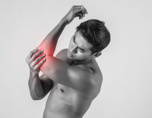
Olecranon bursitis affects the tissue over the posterior point of the elbow. Direct trauma, prolonged leaning on hard surfaces, or repetitive arm extensions can trigger immediate or gradual fluid swelling within the superficial sac.
Prepatellar bursitis, sometimes referred to as housemaid's knee, forms in front of the kneecap. Frequent kneeling on firm flooring causes mechanical microtrauma, triggering inflammation within the tissue sac.
Signs and Symptoms
The clinical presentation of bursitis varies based on anatomical location, underlying cause, and chronicity. Symptoms range from mild localized aching to sharp pain during joint motion.
- Localized Tenderness: Direct pressure on the affected bursa often reproduces sharp pain.
- Swelling and Warmth: Superficial bursae, such as the olecranon or prepatellar bursae, frequently show visible fluid accumulation and local temperature increases.
- Pain During Active Movement: Muscle contractions that compress or stretch the inflamed bursa heighten discomfort.
- Stiffness: Joint motion feels restricted, especially after resting or waking in the morning.
- Sleep Disturbance: Lying directly on an inflamed hip or shoulder frequently worsens discomfort and disrupts sleep cycles.
Mild presentations display localized discomfort only during specific activities or extreme ranges of motion. Severe acute presentations can restrict simple daily functional tasks such as putting on a jacket, rising from a chair, or walking short distances.
Diagnosis
Evaluating bursitis requires a thorough structured clinical assessment combined with diagnostic investigations to rule out alternative mechanical or systemic conditions.
Clinical Assessment
A physical therapist or physician evaluates symptom onset, activity patterns, and daily movement demands. The examination assesses:
- Palpation: Identification of localized points of maximal tenderness over anatomical bursae.
- Range of Motion Testing: Comparison of active and passive joint movements. Active movement typically reproduces pain because surrounding muscles contract and compress the tissue, whereas passive movement may be less painful unless the bursa is compressed at end-range.
- Resisted Strength Testing: Muscle contractions against resistance help differentiate bursitis from primary muscle or tendon tears.
- Functional Movement Screens: Observation of walking mechanics, squatting patterns, or overhead reaching to identify underlying biomechanical contributors.
Clinical special tests, such as shoulder Hawkins-Kennedy testing or hip single-leg stance evaluations, support the clinical impression but do not definitively diagnose tissue changes without clinical synthesis.
Medical Testing
Medical imaging and laboratory tests provide diagnostic clarity when symptoms persist or present atypically:
- Diagnostic Ultrasound: Real-time ultrasound visualizes bursa thickness, fluid volume, and local tissue hypervascularity.
- Magnetic Resonance Imaging (MRI): Detailed soft tissue images differentiate between isolated bursitis, partial tendon tears, bone marrow edema, or joint pathology.
- X-rays: Plain radiography does not show soft tissue bursae directly, but it identifies bony spurs, joint space narrowing, or calcific deposits that contribute to ongoing friction.
- Aspiration and Fluid Analysis: If septic bursitis or crystalline arthropathy such as gout is suspected, a clinician aspirates bursal fluid for laboratory testing, cell counts, and bacterial cultures.
Complications and Risk Factors
Identifying potential risk factors helps tailor intervention strategies and minimize symptom recurrence.
Risk Factors
Modifiable risk factors include excessive training volume, sudden increases in physical activity, poor lifting technique, prolonged static kneeling or leaning, and muscle weakness surrounding major joints. Non-modifiable risk factors involve advancing age, anatomical variations in joint structure, and pre-existing inflammatory joint conditions.
Complications
Unmanaged chronic bursitis can lead to compensatory movement patterns, chronic pain sensitization, secondary muscle weakness, and tendon degradation. When fluid remains trapped under high pressure, surrounding tissues face altered mechanics and elevated stress.
Red Flags
Seek urgent medical evaluation if any of the following symptoms appear:
- Sudden onset of severe redness, warmth, and rapidly spreading skin discoloration over a joint.
- Fever, chills, or systemic illness accompanying joint swelling (signs of septic bursitis).
- Inability to bear weight or complete joint locking.
- Progressive neurological weakness, numbness, or tingling extending down an extremity.
Physical Therapy Exercises
Targeted rehabilitation reduces compression, improves tissue tolerance, and strengthens surrounding musculature to distribute joint loads evenly. Programs progress systematically from symptom management to dynamic functional movements.
Exercise selection must be individualized based on diagnosis, pain severity, age, baseline strength, and movement goals. Stop any exercise that triggers sharp, severe, or rapidly worsening pain.
1. Pendulum Swings (Shoulder Bursitis)
Purpose: Promotes early passive motion and gentle fluid movement in the subacromial space without active muscle contraction.
Starting Position: Lean forward, supporting your non-affected arm on a sturdy table or chair back. Allow the affected arm to dangle straight down toward the floor.
Technique: Gently sway your torso to create momentum, letting your relaxed arm move in small, effortless circles. Complete 15 to 20 gentle circles clockwise and counterclockwise. Avoid active shoulder muscle engagement.
Progression: Increase circle diameter as comfort allows.
Common Mistakes: Using shoulder muscles to pull the arm rather than relying on body momentum.
2. Side-Lying Clamshells (Trochanteric Hip Bursitis)
Purpose: Strengthens gluteus medius and deep hip rotators to stabilize the pelvis during walking and minimize lateral friction over the greater trochanter.
Starting Position: Lie on your non-affected side with knees bent to 90 degrees, hips flexed to 45 degrees, and feet stacked together.
Technique: Keep your feet together while lifting your top knee upward without rotating your hips or lower back backward. Hold the top position for two seconds, then slowly lower. Perform 2 to 3 sets of 10 to 12 repetitions.
Modifications: Place a resistance band around your knees as strength improves.
Common Mistakes: Rolling the top hip backward to lift the leg higher, which recruits hip flexors instead of gluteal muscles.
3. Isometric Straight Leg Raise (Prepatellar Knee Bursitis)
Purpose: Maintains quadriceps strength and knee extension control without compressing the prepatellar bursa through repeated bending.
Starting Position: Lie flat on your back with your unaffected knee bent and foot flat on the floor. Extend your affected knee completely straight.
Technique: Tighten your front thigh muscles, pulling your toes slightly toward your shin. Lift your leg smoothly to the height of the opposite knee. Hold for 3 to 5 seconds, then slowly lower. Perform 2 sets of 10 repetitions.
Modifications: Place a small rolled towel under the knee if full extension causes uncomfortable tension.
Common Mistakes: Arching the lower back or letting the knee bend during the raise.
4. Scapular Retraction and Setting (Shoulder Bursitis)
Purpose: Re-positions the shoulder blade to open the subacromial space, decreasing mechanical pinching of the subacromial bursa.
Starting Position: Sit upright with shoulders relaxed and arms by your sides.
Technique: Draw your shoulder blades downward and gently squeeze them together toward your spine. Hold for 5 seconds without shrugging your neck muscles. Release slowly. Complete 3 sets of 10 repetitions.
Common Mistakes: Arching the lower back or shrugging the shoulders upward toward the ears.
5. Bridging with Pelvic Alignment (Hip and Knee Support)
Purpose: Builds posterior chain strength in gluteal and hamstring muscles to improve hip extension and reduce anterior joint loads.
Starting Position: Lie flat on your back with knees bent, feet hip-width apart and flat on the floor.
Technique: Press through your heels to lift your hips until your thighs align with your torso. Focus on contracting your gluteal muscles. Hold for 3 seconds, then lower smoothly. Perform 3 sets of 10 repetitions.
Common Mistakes: Over-extending the lumbar spine rather than lifting with the hip gluteal muscles.
Medical Treatment
Conservative management serves as the initial approach for non-septic bursitis. Physician-directed options include short-term nonsteroidal anti-inflammatory drugs (NSAIDs) to manage acute pain, temporary bracing, or targeted ice application to minimize swelling.
When severe pain prevents active participation in rehabilitation, a physician may offer a corticosteroid injection directly into the bursal sac. Injections help decrease fluid volume and local inflammation, creating a window of pain relief during which physical therapy exercises can address underlying biomechanical issues. Septic bursitis requires prompt antibiotic therapy and direct medical drainage. In persistent cases that fail conservative care, surgical excision of the chronic bursa (bursectomy) may be considered.
Finding a Physical Therapist
Individualized assessment by a qualified physical therapist helps pinpoint the exact movement deficits contributing to bursal irritation. A therapist creates targeted modification strategies, prescribes appropriate exercise loads, and guides tissue recovery safely.
Schedule an evaluation with a licensed physical therapist to receive a personalized rehabilitation plan tailored to your functional goals.
Medical Disclaimer: The information provided in this article is for educational and informational purposes only and should not be construed as medical advice. Always consult with a licensed physical therapist or healthcare professional before beginning any new exercise or rehabilitation program.
References & Medical Sources
Need Expert Treatment Near You?
Book an appointment with our top-rated physical therapists across twin cities. We bring advanced recovery techniques directly to your neighborhood.
Read Next
Piriformis Syndrome Exercises - HealthandPhysio
A comprehensive, evidence-based guide to physical therapy exercises for piriformis syndrome, designed to relieve deep buttock pain and restore hip mobility.
Cervical Radiculopathy Pain Relief
Evidence-based physical therapy exercises and targeted conservative strategies can significantly reduce pain and nerve irritation caused by cervical radiculopathy.
Fibromyalgia Rehabilitation: A Physical Therapy Guide
Fibromyalgia is a chronic pain condition characterized by widespread musculoskeletal pain, increased sensitivity to normally non-painful or mildly painful stimuli, fatigue, sleep disturbance, and difficulties with physical and cognitive functioning.
Key Insights
Subacromial bursitis is identified in up to 70% of clinical consultations for shoulder pain.
Greater trochanteric pain syndrome affects between 1.8 and 5.4 individuals per 1,000 adults annually.
Conservative treatment, including targeted physical therapy, successfully resolves over 85% of non-septic bursitis cases without surgical intervention.
About the Clinic
Health & Physio focuses on empowering recovery through targeted physical therapy and education. Book an appointment today to start your healing journey.
Find a Specialist