Quick Overview
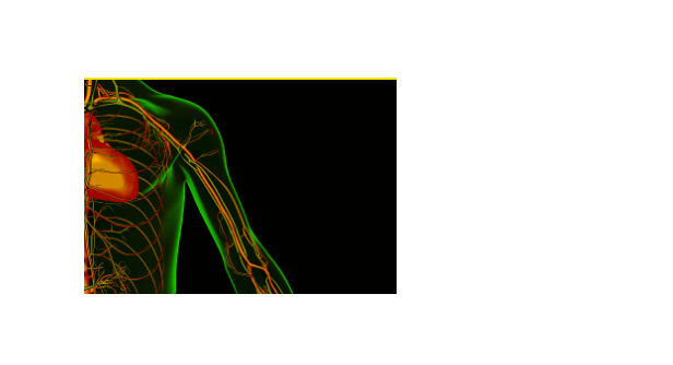
What is axillary vein? The axillary vein is one of the major veins of the upper limb. It conveys blood from the lateral aspect of the thorax, axilla (armpit), and upper limb toward the heart. There is one axillary vein on each side of the body.

Table of Contents
Anatomy
The axillary vein originates at the lower border of the teres major muscle and is a continuation of the basilic vein. It ascends medially through the axilla towards the 1st rib, where it is continued by the subclavian vein. Along its course, it lies anteromedial to the axillary artery, partially overlapping it.
| Vein | Description |
| Origin | Lower border of the teres major muscle |
| Tributaries | Subscapular vein, Circumflex humeral vein, Lateral thoracic veinThoracoacromial vein, Cephalic vein |
| Venous Drainage | Drain blood from the lateral aspect of the thorax, axilla, and upper limb toward the heart. |
Location
This vein is situated in the upper extremity, i,e the axilla (armpit) . It runs from the lower border of the teres major muscle to the 1st rib.
Function
The function of the axillary vein is to drain blood from the lateral aspect of the thorax, axilla, and upper limb toward the heart.
Branches
The tributaries of the AV include:
- Subscapular vein
- Circumflex humeral vein
- Lateral thoracic vein
- Thoracoacromial vein
- Cephalic vein
Clinical Importance
The AV holds significant clinical importance, especially in the context of intravenous access, as it is often utilized for procedures such as central venous catheter insertion. A blockage in the axillary vein can lead to a number of serious problems.
AV Disorders and Treatment
A number of disorders can affect this vein, which include;
- Deep vein thrombosis (DVT): DVT is a blood clot in a deep vein. It can be treated with blood thinners and compression stockings.
- Pulmonary embolism (PE): A PE is a blood clot that travels to the lungs. It can be treated with blood thinners and surgery.
- Axillary vein stenosis: It is the narrowing of the axillary vein. It can be treated with angioplasty and stenting, or surgery.
Prevention of AVD
There are a number of things you can do to prevent AV disorders, including:
- Maintaining a healthy weight
- Exercising regularly
- Avoiding sitting or standing for long periods of time
- Wearing compression stockings if you are at high risk for DVT
Question
1. What is Axillary Vein Thrombosis?
It is a condition characterized by the formation of a blood clot (thrombus) within the AV. It can impede blood flow and potentially lead to swelling, pain, and other symptoms.
2. Axillary Vein Thrombosis Treatment?
Treatment for AV Thrombosis typically involves anticoagulant medications (blood thinners) to prevent further clot formation and facilitate clot dissolution. In some cases, additional interventions may be necessary to remove or break up the clot.
3. How to Perform Axillary Vein Ultrasound?
An AV Ultrasound is a diagnostic procedure performed by a healthcare professional. It involves applying ultrasound gel to the skin overlying the AV and using an ultrasound probe to visualize the vein’s structure and blood flow.
4. Axillary Vein Thrombosis Symptoms?
Symptoms of AV Thrombosis may include swelling, pain, warmth, redness, or tenderness in the affected arm. Some individuals may also experience arm fatigue or a feeling of fullness in the affected area.
5. Blood from the Right Axillary Vein Travels Next to What Vessel?
Blood from the right AV typically travels next to the right Subclavian Artery as it proceeds towards the central venous circulation. The Subclavian Artery is a major arterial vessel in the upper chest.
6. What Are the Two Veins That Merge into the Axillary Vein?
The AV is formed by the convergence of several tributaries, with the two major veins being the Basilic Vein and the Cephalic Vein. These two veins, along with other smaller tributaries, join together to create the Axillary Vein.
