Quick Overview
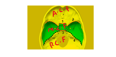
The Middle Cranial Fossa is one of the three cranial fossae, or depressions within the base of the skull, positioned in the middle portion of the cranial cavity. It provides a secure and protective housing for various structures, most notably the temporal lobes of the brain.

Table of Contents
Anatomy of Middle Cranial Fossa
The middle cranial fossa is divided into two parts, i,e the lateral fossa and the medial fossa. The lateral fossa contains the temporal lobe of the brain, while the medial fossa contains the pituitary gland, the sella turcica, and the cavernous sinus.
Location
it is centrally located within the cranial vault. It extends laterally from the lesser wings of the sphenoid bone, continuing towards the temporal bones on either side. This location situates it between the anterior and posterior cranial Fossae.
Read about Cubital fossa : Anatomy, Borders, Contents
Boundaries
It is formed by the following bones:
- Superiorly: Frontal bone
- Medially: Sphenoid bone
- Laterally: Temporal bone
- Inferiorly: Temporal bone and sphenoid bone
Structures
The structures found here are;
- Temporal lobes of the brain
- Pituitary gland
- Sella turcica
- Cavernous sinus
- Internal carotid artery
- Ophthalmic nerve
- Maxillary nerve
- Mandibular nerve
- Trigeminal nerve
- Abducent nerve
- Facial nerve
- Vestibulocochlear nerve
- Greater petrosal nerve
- Lesser petrosal nerve
- Middle meningeal artery
- Ophthalmic artery
- Maxillary artery
- Mandibular artery
Functions
It houses the temporal lobes of the brain, which are responsible for a variety of functions, including;
- Hearing
- Language
- Memory
- Emotion
- Perception
It also houses for the pituitary gland, which is responsible for producing and regulating a variety of hormones, including;
- Growth hormone
- Thyroid-stimulating hormone
- Adrenocorticotropic hormone
- Prolactin
- Luteinizing hormone
- Follicle-stimulating hormone
Clinical Significance
It is susceptible to a number of injuries and conditions, including;
- Head trauma
- Skull fractures
- Brain tumors
- Meningitis
- Aneurysms
- Pituitary tumors
Damage to the structures in can lead to a variety of neurological problems, including;
- Hearing loss
- Speech problems
- Memory loss
- Difficulty thinking
- Vision problems
- Hormonal imbalances
Questions
Q: What are the symptoms of a middle cranial fossa injury?
The symptoms of injury depend on the severity of the injury and the structures that are affected. Common symptoms include:
1- Headache
2- Hearing loss
3- Vision problems
4- Speech problems
5- Memory loss
6- Difficulty thinking
7- Dizziness
8- Nausea and vomiting
9- Weakness or numbness in the face or arms
10- Difficulty with balance and coordination
11- Hormonal imbalances
If you experience any of these symptoms after a head injury, it is important to see a doctor right away.
Q: What are the risk factors for a middle cranial fossa injury?
The risk factors include;
1- Participating in contact sports
2- Engaging in dangerous activities, such as motorcycle riding or rock climbing
3- Falling from a height
4- Being in a car accident
5- Being assaulted
Q: How is a middle cranial fossa injury diagnosed?
The symptoms of a middle cranial fossa injury is typically diagnosed based on a physical examination and the patient’s medical history. Imaging tests, such as a CT scan or MRI, may be ordered to confirm the diagnosis and assess the severity of the injury.
Q: How is a middle cranial fossa injury treated?
The treatment depends on the severity of the injury and the structures that are affected. Mild injuries may be treated with rest, ice, and pain medication. More severe injuries may require surgery.
Q: What is the prognosis for a middle cranial fossa injury?
The prognosis also depends on the severity of the injury and the timeliness of treatment. Some people make a full recovery, while others experience long-term neurological problems.
