Table of Contents
Quick Overview:
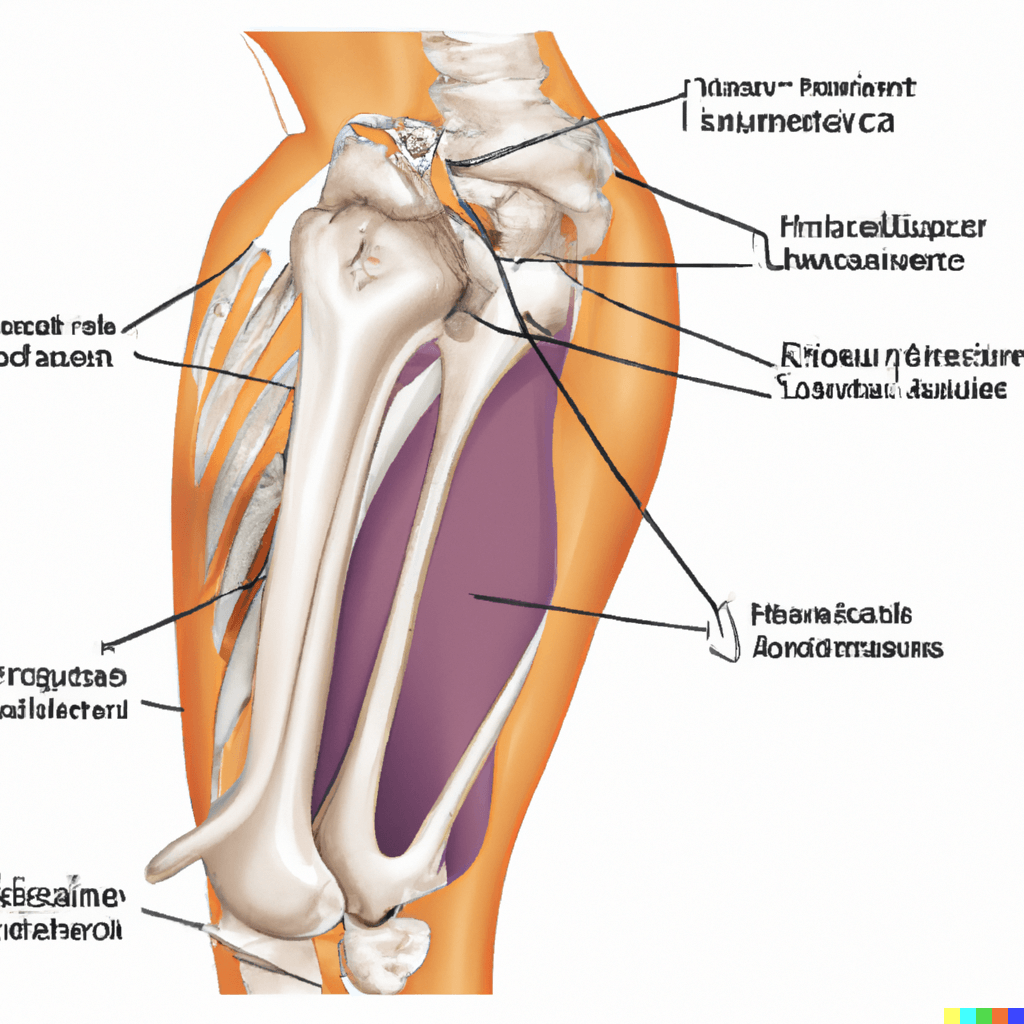
The cubital fossa is an important anatomical region located in the anterior (front) aspect of the elbow, on the distal (lower) end of the humerus bone and the proximal (upper) end of the ulna bone.
It is also known as the elbow pit or antecubital fossa. Specifically, it is a triangular depression or hollow that is bounded by three main structures i,e the brachioradialis muscle on its lateral (outer) side, the pronator teres muscle on the medial (inner) side, and the imaginary line connecting the epicondyles of the humerus bone superiorly (above).
This area is the house of several vital structures, including the brachial artery, the median nerve, and biceps tendon, making it a crucial landmark for medical professionals.
Understanding the anatomy and function of the cubital fossa is essential for diagnosis and treatment of various conditions, such as cubital fossa syndromes and arterial injuries.

Anatomy of the cubital fossa:
The cubital fossa is bounded by three muscles, which are the brachioradialis muscle laterally, the pronator teres muscle medially, the apex of the triangle is formed by the tendon of the biceps brachii muscle.
The base of the triangle is formed by an imaginary line between the epicondyles of the humerus bone. The floor of the cubital fossa is formed by the brachialis muscle.
Borders of the cubital fossa:
Lateral border: Laterally it is formed by the brachioradialis muscle, which runs from the lateral epicondyle of the humerus bone to the styloid process of the radius bone.
Medial border: Medially it is formed by the pronator teres muscle, which runs from the medial epicondyle of the humerus bone to the lateral surface of the radius bone.
Superior border: Superiorly it is formed by an imaginary line connecting the epicondyles of the humerus bone.
Floor: The floor of cubital fossa is formed by the brachialis muscle, which runs from the lower half of the humerus bone to the coronoid process of the ulna bone.
Roof: The roof is formed by the bicipital aponeurosis, fascia, subcutaneous fat and skin
Contents of the cubital fossa:
The cubital fossa contains the following structures:
Brachial artery: Brachial artery is the continuation of the axillary artery. It passes through the cubital fossa and supplies to the forearm.
Median nerve: The median nerve arises from the brachial plexus, runs through the cubital fossa and innervates the muscles of the anterior forearm and the skin of the hand and the fingers.
Radial nerve: This is the largest terminal branch of brachial plexus and passes through the cubital fossa to supply the muscles of the posterior forearm and the skin of the hand.
Biceps tendon: The bicep tendon attaches the biceps muscle to the radius bone of the forearm.
Radial head: This is the proximal end of the radius bone and is located at the lateral aspect of the cubital fossa.
Veins of the cubital fossa:
The cubital fossa contains two important veins i,e the median cubital vein and the cephalic vein. The median cubital vein connects the basilic vein and the cephalic vein, and it is commonly used for blood sampling and intravenous injections. The cephalic vein runs along the lateral aspect of the forearm and drains into the axillary vein.
Cubital fossa syndrome:
Cubital fossa syndrome or Cubital tunnel syndrome is a condition which occurs when there is compression or entrapment of the ulnar nerve at the level of the cubital fossa.
This compression of the ulnar nerve can cause numbness, tingling, and weakness in the hand and forearm. Treatment options for cubital fossa syndrome include conservative management, such as rest and physiotherapy, and surgical intervention in severe cases.
Case Scenario:
Watson, a 35-year-old construction worker, presents to the emergency department with complaints of numbness and tingling in his ring and little fingers. He reports that he has been experiencing these symptoms for the past few weeks, particularly when he is using power tools at work. On examination, there is tenderness over the cubital tunnel in the cubital fossa and positive Tinel’s sign. What condition is John likely to have?
1- Carpal tunnel syndrome
2- Cubital tunnel syndrome
3- Radial tunnel syndrome
4- Median nerve entrapment syndrome
People also ask:
1- What passes through the cubital fossa?
The following structure pass through the cubital fossa;
- Brachial artery
- Median nerve
- Radial nerve
- Biceps tendon
- Brachialis muscle
- Median cubital vein
2- How do you remember the contents of cubital fossa?
The best way to remember the contents of the cubital fossa is to use a mnemonic. Here is the mnemonic through which you can remember cubital fossa structures easily:
“Really Need Beer To Be At My Nicest”
=> Radial nerve
=> Biceps tendon
=> Brachial artery
=> Median nerve
=> Median cubital vein
=> Brachioradialis muscle
=> Anterior ulnar recurrent artery
=> Ulnar nerve
Another mnemonic that can be used is:
“Bread Really Should Buy My New Apple Pie”
=> Brachial artery
=> Radial nerve
=> Median nerve
=> Biceps tendon
=> Median cubital vein
=> Brachioradialis muscle
=> Anterior ulnar recurrent artery
=> Pronator teres muscle
Cubital fossa MCQs:
1- Which muscle forms the floor of the cubital fossa?
A) Brachioradialis
B) Pronator teres
C) Brachialis
D) Triceps
Answer: C) Brachialis
2- What is the main artery of the arm that passes through the cubital fossa?
A) Ulnar artery
B) Radial artery
C) Brachial artery
D) Subclavian artery
Answer: C) Brachial artery
3- Which nerve passes through the cubital tunnel in the cubital fossa?
A) Radial nerve
B) Median nerve
C) Ulnar nerve
D) Musculocutaneous nerve
Answer: C) Ulnar nerve
4- Which vein is commonly used for blood sampling and intravenous injections in the cubital fossa?
A) Radial vein
B) Ulnar vein
C) Brachial vein
D) Median cubital vein
Answer: D) Median cubital vein
5- Which muscle runs from the lateral epicondyle of the humerus to the styloid process of the radius and forms the lateral border of the cubital fossa?
A) Brachioradialis
B) Pronator teres
C) Brachialis
D) Triceps
Answer: A) Brachioradialis
Related Articles
Beck’s Triad: Symptoms, Causes, and Treatment – HealthandPhysio
Hesselbach triangle: Anatomy, Border, Content – HealthandPhysio
