Table of Contents
Quick Overview:
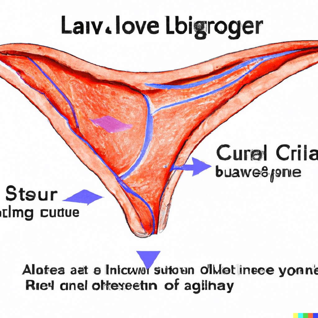
The “Calot’s triangle”, also known as the “cystohepatic triangle”. It is an important anatomical landmark in the body and located in the upper-right quadrant of the abdomen, just beneath the liver. The structures that form the triangle are the common hepatic duct, the cystic duct, and the lower margin of the liver.
This triangle was first described by a French surgeon “Jean-Francois”, in 1891 and named after his name. This triangle is used as an anatomical reference point during laparoscopic cholecystectomy (also known as the surgical removal of gallbladder) and the biliary procedures.
The Calot’s triangle has a great importance due to its anatomy and it contains both the cystic artery and the cystic duct, which supplies blood to the bile and the gallbladder.
It is very important to take care when performing a surgery to avoid any damage to the structures in the Calot’s triangle which can lead to serious complications such as bile duct injury, bleeding and infections.
A better understanding is important about the anatomy and the variations of Calot’s triangle for medical professionals like radiologists, gastroenterologists, and other medical staff who interpret imaging studies of the biliary system and for a successful intervention in the biliary tract.
Calot’s triangle critical view of safety:
Calot’s triangle critical view of safety (CVS) is the technique used by medical professionals performing surgery like laparoscopic cholecystectomy to ensure the safe dissection of the gallbladder from the cystic pedicle without damaging the structures within the triangle.
Using this technique it is important to take a clear view of the cystic duct and the cystic artery before dividing them, and ensure that they are the sole structures being dissected.
The critical view of safety (CVS) technique has three components:
1- The dissection of the gallbladder from the liver bed: The surgeon must make sure that the gallbladder has been completely separated from the liver bed, revealing the cystic plate and the junction of the liver and gallbladder.
2- The Identification of cystic duct and artery: The surgeon must have to identify both the cystic duct and artery and make sure that there are no other structures present in Calot’s triangle that could be confused with them.
3- The Confirmation of the critical view: Once the cystic duct and artery has been identified, the surgeon must confirm that they are the sole structures present in the dissection field. This can be achieved by pushing the gallbladder gently towards the liver bed to obtain a clear view of the triangle.
Boundaries of Calot’s triangle:
The three structures i,e the cystic duct, the common hepatic duct, and the inferior margin of the liver combine and form a triangle-shaped region called “Calot’s triangle”approximately 1-2 cm in size located in the upper-right quadrant of the abdomen, beneath the liver. Followings are structures that form the Calot’s triangle;
1- Cystic duct: The cystic duct from the right border of the triangle and connects the gallbladder to the common bile duct.
2- Common hepatic duct: The common hepatic duct forms the left border of the triangle and is formed by the union of the left and right hepatic ducts, which then drain bile from the liver.
3- Inferior margin of the liver: It forms the superior border of the triangle.
Lymph node in Calot’s triangle:
Lymph nodes are present in this area are a part of the lymphatic drainage system of the biliary tract.The lymphatic vessels that drain the bile ducts and gallbladder run alongside the cystic duct and the common hepatic duct within Calot’s triangle. These lymphatic vessels drain into the lymph nodes that are located in the connective tissue of the triangle.
The lymph nodes present in the Calot’s triangle play an important role in filtering the lymphatic fluid and removing the waste products, bacteria, and the cancer cells that may be present in the bile ducts or gallbladder.
Sometime, the lymph nodes in the triangle may become enlarged due to infection or inflammation of the biliary tract, or due to the presence of cancer cells in the lymphatic system.
When performing surgery, it is important for surgeons to identify the lymph nodes in Calot’s triangle and assess their size and appearance. In some cases the enlarged lymph nodes may indicate the presence of cancer.
Incase of enlarged lymph nodes biopsy may be necessary to confirm a diagnosis of cancer or other pathology. However, care must be taken during lymph node biopsy to avoid any damage to the structures within Calot’s triangle, such as the cystic duct and hepatic artery, that can lead to serious complications.
Name of the lymph nodes in the Calot’s triangle:
The lymph nodes present in Calot’s triangle are called the “cystic duct lymph node” or “the lymph node of Calot“. It may be a single node or a group of nodes that are located near the junction of the cystic duct and the common hepatic duct. The cystic duct lymph node receives lymphatic drainage from the gallbladder, cystic duct, and the common bile duct.
Contents of the Calot’s triangle:
The structure within the Calot’s triangle includes
1- The Cystic duct: The cystic duct connects the gallbladder to the common bile duct and is located on the right side of the triangle.
2- The Cystic artery: The cystic artery is a branch of the right hepatic artery that supplies blood to the gallbladder and is located near the cystic duct.
3- Lymphatics: The lymphatic vessels that drain the bile ducts and gallbladder run alongside the cystic duct and the common hepatic duct within Calot’s triangle. The lymph nodes in this triangle filter the lymphatic fluid and remove waste products, such as bacteria, and cancer cells etc.
4- Connective tissue: The function of the connective tissue within the Calot’s triangle is to hold the structures together and provide support.
Importance of Calot’s triangle:
Calot’s triangle is an important anatomical region with clinical and surgical significance. Having a proper knowledge and better understanding about Calot’s triangle help in the diagnosis and treatment of biliary diseases.
It also helps to ensure the safety and effectiveness of surgical procedures involving the gallbladder. It helps the medical professionals to avoid any damage to the structures within the triangle during the surgery.
Case Scenario about Calot’s triangle:
A 66 years old patient named John presents to the emergency department having a severe abdominal pain and jaundice. The patient also has a past medical history of gallstones and was recently diagnosed with an acute cholecystitis.
After performing CT scan of the abdomen shows a possible cystic duct obstruction. The surgical team decides to perform a laparoscopic cholecystectomy to remove the patient’s gallbladder. Performing the surgery, the surgeon identifies a small lymph node within Calot’s triangle that appears to be enlarged.
Questions;
1- What is the significance of the lymph node in Calot’s triangle?
2- What steps should the surgical team take to ensure the safety of the patient?
MCQs about Calot’s triangle:
1- Which structure forms the upper boundary of Calot’s triangle?
a. Cystic duct
b. Cystic artery
c. Common hepatic duct
d. Inferior margin of the liver
Answer: c. Common hepatic duct
2- What is the function of lymph nodes present within the Calot’s triangle?
a. Filtering lymphatic fluid
b. Producing bile
c. Absorbing nutrients
d. Regulating blood sugar levels
Answer: a. Filtering lymphatic fluid
3- Which artery supplies blood to the gallbladder within the Calot’s triangle?
a. Common hepatic artery
b. Celiac artery
c. Superior mesenteric artery
d. Cystic artery
Answer: d. Cystic artery
4- During a laparoscopic cholecystectomy, which structure within Calot’s triangle must be identified and preserved to prevent damage to the liver?
a. Cystic duct
b. Cystic artery
c. Common hepatic duct
d. Lymph nodes
Answer: b. Cystic artery
5- The conditions which can affect the structures within Calot’s triangle?
a. Acute pancreatitis
b. Diverticulitis
c. Crohn’s disease
d. Gallstones
Answer: d. Gallstones
Related Articles
Beck’s Triad: Symptoms, Causes, and Treatment – HealthandPhysio
Talar Tilt Test: Performance, Diagnosis, Treatment, Importance


I got this web page from my buddy who informed me about
this website and now this time I am visiting this website
and reading very informative articles at this place. https://www.regs.rw/author/enshaa233/
You are so cool! I do not think I’ve truly read through anything like this before.
So great to discover somebody with a few genuine thoughts on this subject matter.
Really.. thanks for starting this up. This web site
is one thing that is needed on the internet, someone with a bit of originality!