Quick Overview
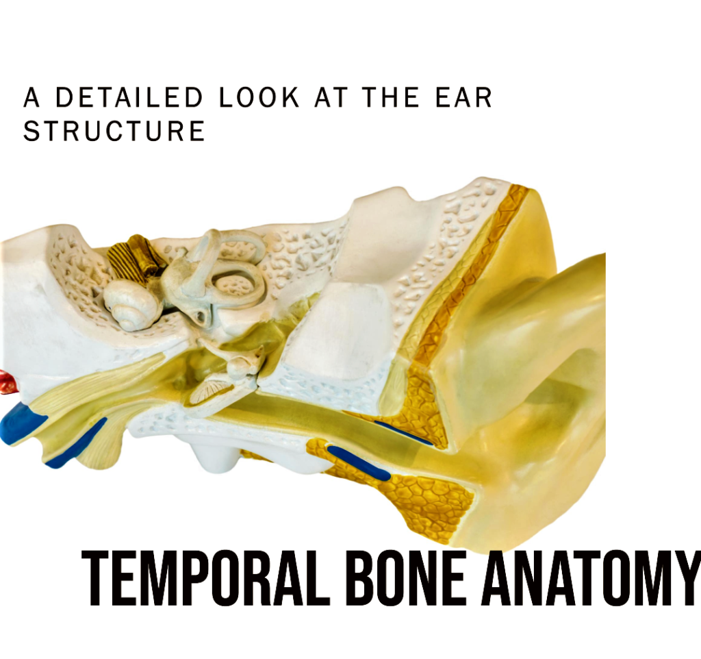
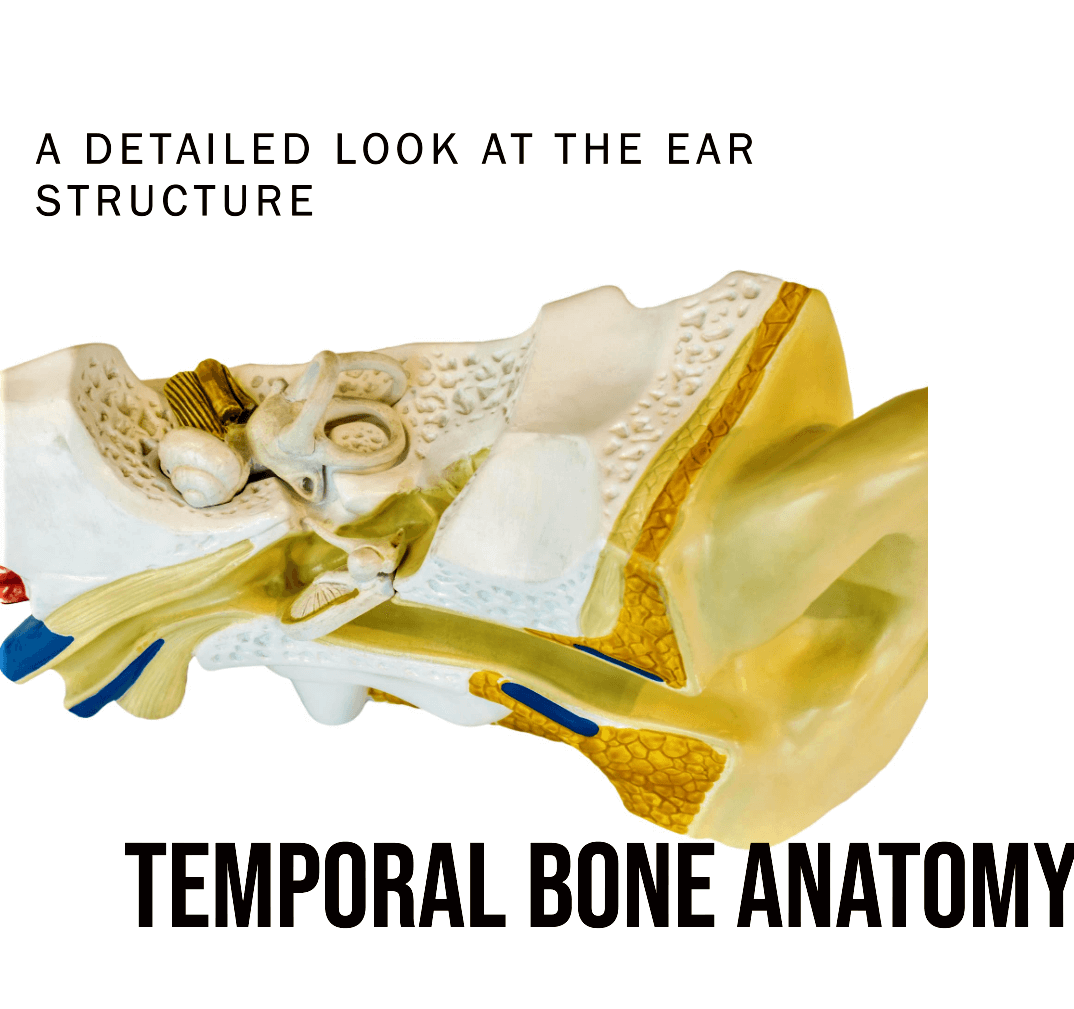
Temporal bones are a pair of bones that form the sides and base of the skull. They are located below the parietal bones and in front of the occipital bone. The temporal bones are important for hearing, balance, and facial expression.
Within the intricate structure of the human skull, the temporal bones stand as guardians of essential sensory organs and the vital anatomical connections.
These bones are located on each side of the skull, i,e the left and right temporal bones play a pivotal role in housing and protecting critical auditory and balancing mechanisms.

Table of Contents
Definition and Location
The temporal bones are a pair of bones situated on each side of the skull, forming the sides and base of the cranium. They are crucial components of the temporal region, housing essential structures related to hearing and equilibrium.
I. Anatomy of Temporal Bones
A. External Features
1. Squamous Portion: The flattened and scale-like part of these bones contributes to the lateral wall of the skull.
2. Zygomatic Process: A bony projection that forms part of the zygomatic arch, which plays a role in facial anatomy.
3. External Acoustic Meatus: A canal-like opening allowing sound waves to travel from the outer ear to the middle ear.
| Feature | Description |
| Squamous Portion | Flattened and scale-like part of the bone |
| Zygomatic Process | Bony projection contributing to zygomatic arch |
| External Acoustic Meatus | Canal-like opening for sound wave transmission |
B. Internal Features
1. Petrous Portion: The dense and pyramid-shaped section of the temporal bone housing the inner ear structures and important cranial nerves.
2. Mastoid Process: It is a bony prominence located behind the ear, and serving as an attachment site for various neck muscles.
3. Styloid Process: A slender, pointed projection which support several ligaments and muscles.
| Feature | Description |
| Petrous Portion | Pyramid-shaped housing inner ear structures |
| Mastoid Process | Prominence for muscle attachment |
| Styloid Process | Slender projection supporting ligaments |
II. Functions
- Hearing: It contain the middle ear, which helps to transmit sound waves from the eardrum to the inner ear.
- Balance: These also contain the inner ear, which helps to maintain balance.
- Chewing: These bones forms the part of the jaw joint, which allows us to chew.
- Facial expressions: With other bones like frontal bone, parietal bones it also contain muscles that help us to make facial expressions.
- Attachment Site for Various Muscles: Several muscles of the head and neck attach to different regions of these bones, facilitating various movements and functions.
III. Developmental and Evolutionary Aspects
A. During Fetal Development
During the fetal development these bones from separate ossification centers which later gradually fusing to form the complete bone.
B. Evolutionary Significance
Throughout human evolution, the temporal bones have undergone adaptations to accommodate changes in cranial capacity and the development of auditory and balance-related structures.
IV. Conditions and Disorders
A. Fractures
Traumatic injuries can lead to bone fractures, which may result in hearing loss, dizziness, and other complications.
B. Temporomandibular Joint (TMJ) Disorders
Issues with the TMJ can cause pain, clicking sounds, and restricted jaw movement, affecting daily activities like eating and speaking.
V. Diagnostic Techniques and Imaging
A. X-rays and CT Scans
X-rays and CT scans are essential diagnostic tools for detecting temporal bone fractures and assessing anatomical structures.
B. MRI Scans
MRI scans provide detailed images, aiding in diagnosing TMJ disorders and other soft tissue conditions.
C. Importance in Audiology
Temporomandibular joint movement can affect hearing, and audiologists consider TMJ health in hearing assessments.
VI. Surgical Procedures
A. Mastoidectomy
Mastoidectomy is a surgical procedure to remove the infected mastoid air cells, often due to chronic ear infections.
B. Cochlear Implants
Cochlear implants, placed in the inner ear during surgery, which assist individuals with severe hearing loss by bypassing damaged cochlear structures.
VII. Facts
- These bones are crucial in estimating the age in forensic investigations, aiding in identifying human remains.
- These bones also have historical significance, often featuring in art and cultural practices throughout human history.
Questions
1. What are temporal bones?
The temporal bones are a pair of bones located on each side of the human skull, forming the sides and base of the cranium.
2. What is the role of temporal bones in the skull structure?
Temporal bones contribute to the structural integrity of the skull, safeguarding delicate brain regions and vital sensory organs.
3. What important structures do temporal bones protect?
Temporal bones house and protect critical structures, including the middle and inner ear, essential for hearing and balance.
4. How do temporal bones articulate with the mandible?
Temporal bones articulate with the mandible, forming the temporomandibular joint (TMJ), enabling jaw movements for speech and chewing.
5. What are common conditions related to temporal bones?
Common conditions include temporal bone fractures, which may lead to hearing loss and dizziness, and temporomandibular joint (TMJ) disorders causing jaw pain and restricted movement.

